Category Archives: Case Profiles

Case Presentation: Utilizing Laser Therapy in the
Treatment of Rasmussen’s Encephalitis

Santiago, R.; Kahn, F.; Kim, S.; Oszarfati, J.

This patient had been diagnosed with Rasmussen’s Encephalitis and decided to independently pursue a course of Laser Therapy, making the decision to select this therapeutic approach prior to surgical intervention which had been advised.

The patient is a 23-year-old female who lives in France and has been diagnosed with Rasmussen’s Encephalitis, characterized by frequent grand mal seizures which she had been experiencing since November 2015. These seizures were poorly controlled by individual or a combination of medications in high dosages. Her condition became more severe in April 2017 when paralysis of the right side of the body developed accompanied by an increase in the severity of her seizures. This was accompanied by pain on the right side of the face and tongue, gradually extending to the right side of the body including both extremities.

A CT scan performed in January 2018 noted a hypodense area in the left parietal region consistent with Chronic Encephalitis. During this period, she began to experience focal seizures of the right lower extremity which was poorly responsive to medication. This prompted treatment with intravenous immunoglobulin therapy over a 3-month period, which improved her condition minimally. Over time she developed decreased muscle tone and lack of coordination of the right side of her body which confined her to a wheelchair, and resulted in increased difficulty writing, eating and functioning on virtually every level. She also began to experience difficulty processing information and developed expressive aphasia. Prior to presenting at our clinic, she had been under the extended care of a Neurologist in France.

Treatment with Laser Therapy was initiated in May 2018 at the Meditech Clinic in Toronto. Therapy began with the application of both large surface arrays utilizing radiation at Red 660 nm and Infrared 840 nm, followed by the Red and Infrared Laser Probes at 660 nm and 830 nm, respectively. Therapy was applied to the cervicothoracic spine region targeting the brainstem, cerebellum and the cervical spine including the cord. Cranial coverage was included following a week of therapy to the cervico-spinal area.

The initial positive response from the patient included an increased range of motion of the right lower extremity, gradually permitting short walks independently. Of significance was the improved quality of her sleep which increased in duration and depth along with emotional stability. This improved her overall status significantly. Her seizures became less severe in frequency and duration and the protocols were adjusted as the various symptomatic improvements continued to improve. The tremors she had experienced in her right leg decreased in severity.

Following two weeks of therapy at the Meditech Clinic in Toronto, the patient noted continuing improvement in her overall well-being, particularly sleep duration and quality and improved range of motion of all affected areas, accompanied by an almost total lack of seizure activity. On that basis, she was provided with a portable unit to continue treatment in France. This followed careful training of the patient and her family who were provided with a variety of protocols to use at home in accordance with symptom changes.

The patient was advised to continue treatment on alternate days including the cervicothoracic spine and the cranium with instructions for change concomitant with her symptomatology. She was advised to communicate with the clinic in Toronto by email for ongoing guidance. She was also advised to continue treatment with IV immunoglobulin therapy and anticonvulsants as indicated by her Neurologist. The patient currently continues to respond to this multifaceted approach and at this time, the proposed cerebral hemispherectomy, which had been previously advised has been cancelled.

Rasmussen’s Encephalitis is a rare and chronic neurological disorder characterized by unilateral hemispheric inflammation of the cerebral cortex, seizures and progressive neurological and cognitive deterioration. At this time, cerebral hemispherectomy is generally offered to patients in this category, particularly those who respond poorly to conventional medication1. Decisions regarding surgical intervention and the appropriate time to institute such measures are challenging to healthcare providers, caregivers and, needless to say, patients, particularly in the absence of a severe neurological deficit and is questionable at best. Immunomodulatory therapy appears to slow rather than halt progression of the disease and does not change the eventual outcome.

Over the past two decades, Laser Therapy has been introduced as an innovative treatment for the modulation of neural activity in order to improve brain function. Treatment requires exposure of the cervical spine and the central nervous system to a low fluence of light using appropriate delivery methods. The safety and ability to customized protocols using Laser Therapy including variations in wavelength, fluence, power density, number and duration of treatments and the mode of application (continuous or pulsed) to the central nervous system have been investigated in many clinical studies. Several reports with regard to the effects of Laser Therapy demonstrate a significant effect on a wide range of CNS disorders2 including epilepsy, traumatic brain injury, neurodegenerative disorders, headaches, vertigo, mobility problems, multiple sclerosis, neuromuscular disorders along with impaired sleep patterns, CVA and transient ischemic episodes.

Although the response of this patient to Laser Therapy may primarily be attributed to its neuromodulatory and neuroprotective effects, the potent anti-inflammatory effect on tissue may have contributed significantly. A number of researchers have demonstrated an increase in adenosine-3’, 5’-cyclic monophosphate (cAMP) following the administration of Laser Therapy. Although it is tempting to suppose that this increase in cAMP is a direct consequence of a rise in ATP following light therapy, clear-cut evidence for this supposition is still beyond the realm of proof. It has been reported that cAMP-elevating agents, i.e. prostaglandin E2, inhibit the synthesis of TNF and therefore downregulated the inflammatory process. Lima et al. investigated the signaling pathways responsible for the anti-inflammatory action of Laser Therapy (administered at 660 nm, 4.5 J cm−2) when applied to the lungs and airways. They found reduced TNF levels in the tissue treated, probably secondary to an increase in cAMP levels3. This would indicate that Laser Therapy may be a useful adjunct in the treatment of certain central nervous system disorders that are accompanied by a significant inflammatory component.

CONCLUSION:
This case of Rasmussen’s Chronic Encephalitis serves as an example of how Laser Therapy can be utilized in the neuromodulation of a serious brain disorder and demonstrates how Laser Therapy can potentially be a significant factor in the neuromodulation of both the central and peripheral nervous system where conventional therapies do not offer solutions. This is based on a significant improvement in this patient’s overall status and clearly illustrates the potentials of Laser Therapy in the treatment of these conditions. This is largely due to its neuromodulatory, neuroprotective and anti-inflammatory effects and supports ongoing research and application of this treatment for this and other neurological conditions. During a relatively brief course of treatment which is continuing, the majority of the patient’s symptoms were markedly reduced and functions at most levels have improved substantially. Currently as her treatment is effective, surgery need not be considered. Moreover, the patient and her family are pleased with the positive changes noted.

  1. Varadkar S, Bien C, Kruse C, Jensen F, Bauer F, Pardo C, Vincent A, Mathern G, Cross JH. Rasmussen’s encephalitis: clinical features, pathobiology, and treatment advances. Lancet Neurol. 2014 Feb; 13(2): 195–205.
  2. Salehpour F, Mahmoudi J, Kamari F, Sadigh-Eteghad S, Rasta SH, Hamblin M. Brain Photobiomodulation Therapy: a Narrative Review. Molecular Neurobiology https://doi.org/10.1007/s12035-017-0852-4.
  3. Freitas LF, Hamblin M. Proposed Mechanisms of Photobiomodulation or Low-Level Light Therapy. IEEE J Sel Top Quantum Electron. 2016 May-Jun; 22(3): 7000417.

A Summary of the Signs and Symptoms of Cerebral Concussion and Guidance – Re: Clinical Management

Etiological factors may vary from a minor blow to the head, often combined with a whiplash-type injury of the cervical spine resulting in a symptom complex that may be minimal in degree. However, when the trauma is more severe, it may render the patient comatose for prolonged periods of time and even result in death. In most instances, symptoms are brief in duration and last for a matter of days to several months.

The Center for Disease Control and Prevention defines concussion as “a complex pathophysiological process affecting the brain, induced by traumatic biomechanical forces, secondary to direct or indirect forces applied to the cranium.”

Symptoms can last from minutes to days and in some instances become chronic or permanent. Early symptoms include headaches, pain in the cervical spine, nausea, dizziness, an inability to focus or concentrate, memory loss, visual disturbances and in a small number of cases induce loss of consciousness or even coma.

The Glasgow Coma Scale classifies concussion based upon the level of consciousness of the patient. Mild concussion typically allows complete neurological recovery. Moderate concussion can result in stupor and lethargy. In cases of severe concussion, patients may experience a comatose state, a heightened risk of hypotension, hypoxemia and edema of the brain. Invariably, all forms of concussion result in varying degrees of inflammation of neurological tissue, accompanied by pain and neurological impairment.

It has been determined that MRI, PET scans, EEG studies, etc., generally do not reveal any significant abnormalities in the early phases of the disease. Macroscopic changes of cellular tissues may not accompany the symptoms that exist until a significant period of time has elapsed.

Usually, traumatic episodes that result in concussion include a collision with an opposing player when engaged in sports activities, falls and other incidents sustained in the home, motor vehicle accidents and the numerous traumatic episodes related to military activities. The majority of episodes cause only minor symptoms, which usually disappear over a finite period of time, in most cases without any specific therapy being applied.

It has been calculated that somewhere in the vicinity of 10 to 20% of all cerebral concussions that have been formally diagnosed result in symptoms, which if they persist over a period of time may become chronic. In all of these cases, appropriate therapy should be applied and continued until the patient has been asymptomatic for a period of at least 2 months while engaged in relatively normal activities.

The symptoms most frequently reported consist of headaches which may take many forms. Generally, they are fronto-occipital in location accompanied by pressure sensations, stabbing, aching, throbbing, etc. They may be intermittent or exist on a 24/7 basis. The majority of these patients also complain of pain in the cervical spine with or without radiation to the upper extremities. Insomnia, irregular sleep patterns and cognitive impediments may be minimal to extreme. These include difficulty in finding words, the normal comprehension of reading material, TV content, an inability to focus and concentrate and in essence, mentation is impaired in varying degrees. Memory loss may be short or long term and in some instances both. Often this relates to the duration of the period of loss of consciousness, which however in the majority of cases does not occur. As previously indicated, the injury may induce a prolonged state of coma or even death.

Symptoms may also include a loss of sense of taste, reduced auditory acuity, tinnitus and visual disturbances including blurring, double vision, etc. Both light and sound sensitivity are frequent and may persist. The patient may be unable to attend school, work, lose the ability to socialize and be unable to engage in normal relationships. Irritability with periods of labile mood disorders may be accompanied by loss of anger control. Fatigue, anxiety and depression may be present in both the early and late phase of cerebral concussion. With the development of chronicity however, they may become the most dominant long-term problems.

An increasing number of neuroscientists believe that many years later, neurological diseases such as Parkinson’s disease and the dementias may result from a traumatic episode of the brain experienced much earlier in life and long forgotten.

All patients who have sustained cerebral concussion with persistent symptoms should be monitored until asymptomatic and in most cases based on clinical findings and progress, should be subjected to curative Laser Therapy and in some instances preventative treatment.

Certainly, in patients who remain symptomatic even for a week without improvement, a course of Laser Therapy based on thorough clinical evaluation is advisable. A standardized or individual customized course of treatment for each patient should be administered as clinically indicated.

In conclusion, all patients who have sustained a cerebral concussion should undergo an ongoing course of Laser Therapy, unless symptoms improve rapidly post-trauma spontaneously. The purpose for this is not only to relieve symptoms but to prevent chronic damage to the intracranial tissues. Therapy can be standardized or customized and ideally should be applied every 2 days initially.
• Depending on clinical change, the protocols are changed according to the progression or resolution of the severity of the symptoms.
• This applies particularly for patients who are severely affected and present for an assessment relatively early after the injury.
• In chronic cases where symptoms persist regardless of the time period elapsed since the trauma, curative therapy should continue for at least 2 months subsequent to the resolution of all symptoms. Depending on the nature and duration of the injury and unless symptoms are improving rapidly spontaneously, treatment should be applied as early as possible and continued for a minimum of 8 weeks during which the patient is able to engage in normal activities.
• Each case must be assessed on its own merits and treated accordingly.
• In our experience, Laser Therapy should be considered the treatment of choice and be continued until the patient is asymptomatic, fully active and does not require any other therapy, including medications.

Conclusion: All patients who have sustained a cerebral concussion and continue to demonstrate symptoms receive a course of Laser Therapy tailored to their individual situation. Ideally therapy should be continued every second day until the patient has become asymptomatic for at least 2 months.

Generally, protocol settings are initiated at lower levels and increased in accordance with clinical change. As symptoms diminish in the degree of severity, the frequency of treatment can be gradually reduced, but not during the first 2 to 4 weeks.

All patients require periodic clinical assessment incorporating appropriate measurements to determine improvement as the course of treatment progresses. Symptoms should diminish rapidly in most instances when all appropriate parameters are observed.

The entire process should result in a significant improvement/cure rate in all categories of cases treated. This should be accompanied by the restoration of the patient’s quality of life, normal activity levels, and a total absence of symptoms.

BioFlex Laser Therapy Patient Review – Agnese Nunno

I came to Dr. Kahn because I wanted help with my long term memory. I noticed slowly over time, that I had started to forget events that happened in my life, words of songs I use to sing every day amongst other things. I had no idea how much the laser therapy could help heal, amongst many other things.

After the first two treatments I started to notice small changes occur. My sensitivity to light started to become less pronounced, as well as my sensitivity to sound. This was incredibly encouraging to me as I did not think changes would happen so quickly, nor those kinds.

On my own time, throughout the laser therapy treatments, I would work to rewire my brain. I paid close attention to things that occurred that were out of the norm for me. For example, one day while I Was eating, I noticed that I absentmindedly picked up my fork with my non dominant hand and started to eat with ease. The change that become most pronounced throughout the treatments was my overall mood was calmer and happier making my thoughts clearer. My concentration on topics often deemed less enjoyable, became easier to the point where I would describe it as not human. I became able to focus with so much strength that hours would go by before I realized how much time had actually passed. I became able to translate my thoughts into words faster, with the uncategorized fog I had lived with for so many years in my head, disappearing. My procrastination habits vanished where beginning topics no longer needed my rituals of the slow staggered start with scrolling through Facebook, watching YouTube videos until I’ve psyched myself up enough to begin.

I would read different articles and parts of books on rewiring the brain, and apply what I could. I would practice memorizing new topics using all the available senses. I looked at the laser therapy as a tool to work with and I used it as such.

My random anxiety where I would be performing a task and out of the blue have a panic attack for no reason, went from being a 10/10 to a 0/10. My depression and random crying had ceased to exist where now any reason I may feel down or anxious can be directly explained to an event occurring or that has happened. Same can be said for my random unexplained feeling of irritability, which went from again being a 10/10 to a 0/10.

Interesting to note, in order to control my anxiety I had slowly progressed to drinking every night more than a healthy amount for the past five years, which helped calm me but was obviously becoming a problem. The laser therapy had made the desire to drink become less pronounced, and I took that clutch to help me stop drinking.
I sleep sounder at night, and I’m less tiered than I was before where it felt like no amount of sleep was enough. I find that it is also easier to fall asleep without having to take any sleep aids like melatonin.

When in an argument or discussion I find I can now keep up, as before it was difficult because due to the stress I would forget what I had just said or what the other person has just said. As a result this has given me confidence which has helped me not to be afraid as I previously was. The laser therapy has helped me in other areas not expected as well, but the ones that I have spoken about I thought were worth mentioning.

Overall I am incredibly happy with the results of the laser therapy. Currently I’ve had over 15 laser treatments and I would say I feel 98% healed. The 2% that remains is my noise sensitivity, which I have been able to narrow down to two specific situations that affect me. First one being, when I’m on a crowded subway during rush hour where it becomes stressful due to the crowds, the pushing, touching, shoving etc., and the other being people yelling. I feel that the reason why I’m not 100% healed yet is because right after my treatment I have to take the subway back home. I compare it to, having just had a foot cast taken off and then running a marathon right away. I still live at home with my 5 younger siblings with four of them being teenagers and there tends to be a lot of arguments and yelling between them which will make my ears start to ring and vibrate. In the end though, the reason I came to Dr. Kahn was for my long term memory and as far as I can see it has taken care of that concern.

Abstract – Case History – Cerebral Concussion

This 28-year-old bank employee sustained at least three concussions and injuries to the cervical spine, the first one occurring at age 10 years. The concussions were invariably activity/sports related and were not accompanied by any episodes of loss of consciousness.

As the number of traumatic episodes increased, she began to develop symptoms which early on were sometimes relieved by craniosacral massage but as symptoms progressed, they became chronic.

Over the years, she developed the following symptoms: severe long term memory loss and fronto-temporal headaches characterized by extreme pressure sensations, which could be quite debilitating. Sleep became increasingly problematic, she began to suffer from light sensitivity, anxiety, depression, fatigue, tinnitus and an inability to concentrate. From the sixth grade on, she also developed a learning disability, which proved to be a significant impediment.

Symptoms prior to attending the Meditech Laser Therapy Clinic in January 2018 had limited both physical and mental activities and diminished her quality of life.

The patient’s testimonial included in this abstract clearly relates her story subsequent to undergoing a course of BioFlex Laser Therapy.

During the course of this year at Meditech, the clinic will treat approximately one thousand similar cases. Once again, this patient demonstrates the high level of effectiveness of Laser Technology in treating the majority of patients suffering from this disease entity.

Clinical Abstract

Purpose of Article – to Demonstrate the Efficacy of Laser Therapy in the Treatment of Neurological Problems.

The case presented is that of a 62-year-old Firefighter. The patient began to experience unilateral sensorineural hearing loss in the left ear, along with vertigo and severe headaches in April, 2016. An MRI of the brain was performed a week later and revealed that a grade 2 ependymoma was present in the 4th ventricle. This was impinging on the dorsal medulla.

The patient was subjected to a craniotomy in order to remove the tumour on June 7, 2016, followed by a course of radiation therapy.

During his recovery, he developed paralysis of the left lateral rectus muscle leading to diplopia. In addition, he was experiencing a lack of balance, nystagmus, severe headaches, slurred speech, difficulty swallowing and fatigue.
Prism glasses were prescribed to alleviate the visual problem, but no improvement was noted. He was told to wait 18 months in the hope that the diplopia, etc. would improve. 15 months post-surgery, his symptoms persisted and some additional symptoms had developed. These included sleep disturbances, memory loss, irritability, depression, anxiety, loss of taste and tinnitus. He presented at the Meditech Rehabilitation Clinic on January 9, 2018.

Physical Examination:

The patient was noted to be right-handed. The right grip was 105 lbs. and the left was 100 lbs.
Range of motion of the cervical spine with regard to flexion, extension, lateral rotation and lateral flexion was 50% of normal.
There was a loss of the normal curvature of the cervical spine and moderate tenderness, C1-C6.

There was limited abduction of the left lateral rectus muscle with bilateral nystagmus on gazing to the left.

Initial Diagnosis:
Post-Surgical Resection of Malignant Ependymoma
4th Ventricle with Multiple Neurological Sequelae.

TREATMENT AND PROGRESS:

The patient initiated treatment using Laser Therapy at the Meditech clinic on January 9, 2018. Initially, treatment was confined to the occiput, cervical and upper thoracic spine. Irradiation of the brainstem and cerebellum were included with the cervical treatments. Following 5 treatments of these areas over 5 days, treatments were extended to the cerebral hemispheres, initially targeting the occipital lobe. Over several additional days, treatment of the cranial hemispheres was extended to include the temporo-parietal and the frontal lobes. These targeted the motor, sensory and speech centres of the brain.

After 3 sessions of daily treatment at our clinic, including the cerebral hemispheres, the patient noted that his horizontal gaze was more balanced, and the diplopia from which he suffered was disappearing. After an additional week, there was marked improvement in the lateral abduction of his left eye and the double vision had disappeared. His energy levels were elevated and he was able to be more active physically. At this point, his caregiver had been trained to utilize the equipment preset for treatment at home and this is continuing.

RELEVANT DATA:

Ependymomas are a type of tumour of the glial cells (supporting cells of the brain) that starts in the ependyma. The ependyma lines the fluid-filled spaces in the ventricles (cavities) of the brain and the centre of the spinal cord. Ependymomas spread to the cerebrospinal fluid more often than other gliomas. They do not spread outside the brain or spinal cord. Based on the World Health Organization (WHO) grading system, ependymomas are classified as low to high grade (grades 1–3). Low-grade tumours (grade 1) are more common in the spine while grade 2 tumours are more common in the brain.

Symptoms depend on the tumor location. The most frequent symptoms include:
• Headache and intracranial pressure
• Nausea and/or vomiting
• Blurred vision
• Weakness or numbness and tingling
The cause is not known. It rarely occurs in multiple family members, although does occur in people with Neurofibromatosis type 21.

Treatment is usually based on the size of the tumour and the symptoms that occur. Surgery may be performed to remove all of the tumour or as much of the mass as possible. Low-grade tumours may not need further treatment if the entire tumour can be removed. Radiation therapy may be given after surgery for grade 1 and 2 tumours if the tumours can’t be completely removed. Radiation therapy is given after surgery for anaplastic tumours. It may also be used to treat tumours that return after treatment (called recurrent ependymomas) if radiation therapy was not given previously. It may be given to the entire brain and spinal cord if tests show that the cancer has spread through the cerebrospinal fluid2.

Brain surgery and the consequent radiation therapy following the procedure is a major medical event which carries numerous associated risks including an allergic reaction to anesthesia, intracranial bleeding, blood clots, edema, coma, impaired speech, vision, coordination, or balance, infection in the brain or at the wound site, memory problems, seizures and strokes. Aside from some of the side effects mentioned, our patient also sustained nerve damage of the abducens nerve (6th cranial nerve)3.

The sixth nerve leaves the brainstem at the pontomedullary junction and follows an upward and outward path. The long intracranial course of the abducens nerve makes it vulnerable to injuries, most often at the site of the dural entry point and at the petrous apex. Although traumatic palsy of the sixth nerve is usually seen in combination with intracranial hemorrhage, skull fracture, facial fracture or elevated intracranial pressure, it may occur in the absence of such lesions.

The appropriate management of posttraumatic or post-surgical isolated abducens nerve palsy remains a matter of debate. Spontaneous recovery from sixth nerve palsy usually occurs within 6 months, with improvement noted in most cases at 3 months4. Our patient remained symptomatic 15 months post-surgery.

Laser therapy has a favourable prognosis in the regeneration of peripheral nerves in both neurosensory and neuromotor deficits, such as trigeminal neuralgia, neuropathy, low back pain with sciatica, and herpes zoster. Application of laser produces both local and systemic effects that can stimulate the nerve regeneration process. Moreover, laser therapy improves the recovery of the injured peripheral nerve and decreases post-traumatic retrograde degeneration of the neurons in the corresponding segments of the spinal cord. Research studies have shown that Laser therapy increases the functional activity of the injured peripheral nerve, prevents or decreases degeneration in corresponding motor neurons of the spinal cord, and improves axonal growth and myelinisation.

Bernal previously found that Laser Therapy is an excellent complementary medium for the recovery of facial nerve paralysis and provides a painless therapeutic alternative without side effects that can be used on any type of patient, including those who cannot use corticosteroids, such as diabetics and hypertensive patients. In addition, Ladalardo et al. studied the effect of GaAs diode laser in patients with Bell’s palsy and used THE House-Brackman Facial Nerve Grading System (HBS) to assess the outcome. In that study, patients who received the treatment showed a functional improvement ranging between one and three grades on the HBS. One of the possible explanations of the laser effect is through an increase in the activity of enzymes involved in the mitochondrial respiratory chain, such as cytochrome C oxidase and adenosine triphosphatase (ATP), thereby leading to an increase in ATP production in mitochondria. In addition, it increases DNA synthesis as well as collagen and pro-collagen production. The anti-inflammatory effect of laser therapy can be caused by a reduction in the levels of pro-inflammatory cytokines, such as interleukin-1 alpha (IL-1α) and IL-1 beta (IL-1β) as well as an increase in the levels of anti-inflammatory growth factors and cytokines, such as basic fibroblast growth factor (bFGF), platelet-derived growth factor (PDGF), and transforming growth factor-beta (TGF-β). In addition, laser irradiation causes dilatation of blood vessels, which also leads to a reduction in swelling caused by inflammation. It may also have inhibitory effects on the release of prostaglandins, cytokine levels, and cyclooxygenase (Cox) 2, and has been shown to accelerate cell proliferation, collagen synthesis, and tissue repair. Laser Therapy may also have a direct effect on nerve structures, which could increase the speed of recovery of the conduction block or inhibit Aδ and C fiber transmission. Moreover, it was reported that
Laser Therapy significantly widens the arterial and capillary vessels, increases
microcirculation, activates angiogenesis and reduces edema caused by inflammation. It also stimulates the immunological process and nerve regeneration5.

Our experience in the treatment of this patient firmly establishes the positive effects of Laser Therapy. Based on current therapeutic methods, this approach is superior to those being utilized and can therefore be termed the treatment of choice in the resolution of symptoms associated with these neurological conditions. Moreover, application is safe and does not have any of the potential adverse effects of steroids, botulinum toxin injections and unquestionably accelerates the resolution of post-surgical sequelae.


References:

1. http://www.cancer.ca/en/cancer-information/cancer-type/brain-spinal/brain-and-spinal-tumours/ependymoma/?region=on

2. http://www.cern-foundation.org/education/ependymoma-basics

3. https://www.healthline.com/health/brain-surgery#followup

4. Asproudis I, Vourda E, Zafeiropoulos P, Katsanos A, and Tzoufi M. Isolated abducens nerve palsy after closed head injury in a child. Oman J Ophthalmol. 2015 Sep-Dec; 8(3): 179–180.

5. Salaheldien M, Alayat M, Elsodany AM, Abdel Raouf El Fiky A. Efficacy of high and low level laser therapy in the treatment of Bell’s palsy: A randomized double blind placebo controlled trial. Lasers Med Sci (2014) 29:335–342

Clinical Abstract

Edit: February 6, 2018

CLINICAL ABSTRACT

Purpose of Article: To demonstrate the efficacy of Laser Therapy in the Treatment of Chronic Cerebral Concussion.

The patient is a 38-year-old individual who presented complaining of multiple cognitive impediments. On careful questioning, he recalled sustaining a severe concussion at age 5, after falling six feet from a fence. Evidently he lost consciousness for an undetermined period of time and was taken to the hospital by his parents. A number of investigative procedures were performed, however no therapy was provided.

From that time on, the patient had a difficult upbringing, experiencing problems with all relationships including his parents, at school working with teachers and subsequent to completing his education, maintaining a job. He had been on Zoloft for depression for many years. He stated that he had been diagnosed with “Obsessive Compulsive Disorder”, has difficulty organizing, is subject to chronic “brain fog” and has severe pain in the cervical spine. These problems had become well established and the patient summarized his status as being unable to get along with anyone, achieve any level of success in his work objectives and generally preferred to live alone as an introverted, problematic recluse.

PHYSICAL EXAMINATION:
On initial examination on August 18, 2015, the patient was noted to be right-handed. The right grip was 90 lbs and the left was 80 lb. Range of motion of the cervical spine with regard to flexion, extension, lateral rotation and lateral flexion was 60% of normal. A loss of the normal curvature of the cervical spine was noted, along with significant para-cervical muscle spasm and moderate tenderness from the occiput to T1.

The patient’s attitude was extremely negative and with regard to movements and speech, he appeared to be hypoactive. The neurological examination was grossly within normal limits however a bony depression was noted over the left frontal bone, attesting to the initial injury at age 5 years. Generally speaking, the patient was minimally communicative and appeared to be locked in a protective shell.

The initial diagnosis formulated after a prolonged period of questioning, and the physical examination, was–Cerebral Concussion (chronic, severe).

The case clearly represented the clinical picture of a chronic case of Cerebral Concussion and it was somewhat doubtful after the period of time that had elapsed from his initial injury that improvement in his condition could be provided.

COURSE OF TREATMENT: Treatment was initiated using the BioFlex Laser Therapy System on August 22, 2015. Initially, treatment was confined to the cervical spine, including the occiput and upper thoracic spine. Irradiation of the brainstem and cerebellum were included in this process. Following five treatments of this area on consecutive days, treatments were extended to the cerebral hemispheres. Initially only the occipital lobes were targeted but as the course of treatment advanced, treatments were extended to the temporo-parietal and frontal lobes. These focused on the motor, sensory and speech centres of the brain.

After the initial three days of treatment, the patient reported that he was experiencing petit mal seizures at night. He also revealed that the house he grew up in had high lead levels which he was exposed to during his formative years. At this point in the course of treatment, he noted that the chronic “brain fog” was gradually being replaced by clarity and his reading abilities had begun to improve.

At this time, he began to reduce the intake of Zoloft, which had been prescribed for his chronic depression.

As therapy progressed, range of motion of the cervical spine improved to a relatively normal level. After 12 treatments of the cervical spine over a course of three weeks, along with applications to the cerebral hemispheres, he reported that he was able to engage in more physical activities and his mental clarity continued to improve. In addition, he commented that his emotional status was more stable, his energy levels had increased and the periodic petit mal seizures had almost completely disappeared. After six weeks of treatment, the patient felt that he had improved sufficiently on the functional level that he decided to discontinue treatments.

Two years following the completion of treatment, the patient presented with another problem and at that time, we did not recognize him. There was a complete change in his appearance, demeanor and his communicative skills. On questioning, the patient revealed that his lifestyle had improved substantially, although he was still using Zoloft periodically. Conversational levels were relatively normal, the patient was optimistic and there was no evidence of any psychological or behavioural problems.

He indicated that he had been holding a steady job for the past year, had moved to a higher level environment, appeared to be well groomed and communicated with a great deal of self-assurance. Indeed, when I saw him in the waiting room, I had difficulty recognizing him. For the first time since I had known him, he was optimistic with regard to the future and felt that his life was continuing to improve on all levels.

RELEVANT COMMENTS: In the past, there has been minimal progress in developing effective treatments for chronic brain injuries, particularly in cases of multiple cerebral Concussions. This patient illustrated the tremendous benefit that can be induced by appropriate Laser Therapy. The changes in this patient were dramatic from all perspectives, particularly his speech and energy levels. The patient stated that the quality of his life had normalized and looked forward to the future with optimism.

CLINICAL ABSTRACT –
Fred Kahn, MD, FRCS(C), Ronaldo Santiago, MD

PURPOSE OF ARTICLE
To Illustrate The Efficacy Of Light Therapy In The Treatment Of Neurological Pathologies.

PATIENT PROFILE:

Currently he is under the care of his family physician, a neurologist, urologist, etc.

He sustained his initial concussion as a child when he fell out of a tree and lost consciousness for an undetermined period of time. A severe whiplash injury occurred in his 20s, accompanied by trauma to the cranium and three years ago, he had another concussion (moderate/severe) when he sustained a blow to the head at work.

He has been on Acyclovir for the past six months, presumably for treatment of Epstein-Barr Syndrome. Symptoms over the past two years persist without relief and include severe dizziness, headaches, insomnia, fatigue and a complete inability to function at all levels. The patient has been unable to work, is relatively immobile, unable to speak and relates a host of additional cognitive symptoms. He has been subjected to ongoing physiotherapy, occupational therapy, speech therapy, numerous specialist consultants and the employment of an extensive variety of therapeutic options, none of which have improved his status.

PHYSICAL EXAMINATION NOVEMBER 19, 2017:

  • The patient moves slowly and has a shuffling gait.
  • His facial expressions are relatively rigid.
  • Despite his current age of 52 years, his appearance and movements resemble someone in their 80s.
  • The patient is right-handed. The right grip is 50 lbs and the left 40 lbs.
  • Lateral abduction of both shoulders is to 90° only and all movements of both shoulders are restricted in varying degrees.
  • Range of motion of the cervical spine with regard to flexion, extension, lateral rotation and lateral flexion is less than 20% of normal and a similar range of motion of the thoracolumbar spine is 25% of normal.
  • Straight leg raising is 50° bilaterally.
  • Range of motion of both hips is minimal.
  • A reverse thoracolumbar scoliosis with the lumbar apex to the right is noted to be present.
  • Varying degrees of tenderness exist over the cervical, thoracic and lumbar spine.
  • The patient speaks slowly and his voice demonstrates minimal volume.
  • He lacks any affect.
  • His speech is hesitant and the sounds are almost unintelligible.
  • His wife acts as an interpreter which is helpful.

INITIAL DIAGNOSIS:

  1. Neurodegenerative Disorder (Multiple System Atrophy)
  2. Parkinson’s Disease.

TREATMENT AT MEDITECH CLINIC:

The patient commenced treatment with Laser Therapy at our clinic, and this was applied over the cervical, thoracic and lumbar spine, along with both shoulder joints. After five treatments to these areas, therapy was extended to the cerebral hemispheres, initially targeting the occipital lobe and eventually including the temporo-parietal and the frontal lobe

After seven days of daily treatment, there was noticeable improvement in his gait. The patient was able to rise from the seated position without help and recovered a relatively normal gait. His sleep problem was completely resolved and he was able to smile and speak in a normal fashion. Comprehension of all verbal communications had been largely restored.

DISCUSSION:

Multiple system atrophy (MSA) is a rare, progressive neurodegenerative disorder characterized by a combination of symptoms that affect both the autonomic nervous system (the part of the nervous system that controls involuntary action such as blood pressure or digestion) and movement. The symptoms reflect the progressive loss of function and apoptosis of nerve cells in the central nervous system.

Autonomic failure symptoms include fainting spells and problems with heart rate, erectile dysfunction and bladder control. Motor impairments (loss of or limited muscle control or movement, or limited mobility) may include tremor, rigidity, and/or loss of muscle coordination as well as difficulties with speech and gait. As these features are similar to Parkinson’s disease, it may be difficult to distinguish these disorders early in the course of the disease.

MSA can be divided into two different types:

  1. the Parkinsonian type (MSA-P), with primary characteristics similar to Parkinson’s disease (such as moving slowly, stiffness, and tremor) along with problems of balance, coordination, and autonomic nervous system dysfunction, and;
  2. the Cerebellar type (MSA-C), with primary symptoms featuring ataxia (problems with balance and coordination), difficulty swallowing, speech abnormalities or a quavering voice, and abnormal eye movements (“cerebellar” reflects a part of the brain involved with coordination)

The cause of MSA is unknown. There is noted accumulation of the protein alpha-synuclein in glial cells that support nerve cells in the brain, primarily oligodendroglia which produces myelin in the central nervous system. The same protein also accumulates in Parkinson’s disease, but within the nerve cells itself as opposed to the supporting glial cells in MSA1.

There are no known treatments to delay the progressive neurodegeneration associated with MSA and at this time there is no known cure. Current treatments are mostly supportive and convey minimal clinical benefit. In this case, the patient was referred to physiotherapists, occupational therapists, acupuncture practitioners, and an integrative medicine practitioner who detected the Epstein-Barr viral (EBV) antigen in his system. Presumably this is the reason he was placed on Acyclovir, which he felt did not convey any benefit.

Currently there are no studies that mention a connection between MSA and EBV, however the latter has been linked to an increased potential to develop Multiple Sclerosis (MS). For some individuals, Levodopa may improve motor function, however improvement is minimal and diminishes as the disease progresses.

Using cell models of MSA, scientists were able to show that both damage to the mitochondria (cellular “power plants”) and the generation of abnormal alpha-synuclein aggregates may contribute to the development of MSA. In a study conducted by Blin, et al, a significant age-related decrease in the activity of mitochondrial respiratory chain complex I was observed, supporting the hypothesis of a wide-spread mitochondrial complex I deficiency in PD and MSA as compared to age-matched controls, who only showed age-related deficiency. Deficits in complex III and IV activity (which includes cytochrome c oxidase) was also observed, but which was restricted to a few patients2.

Many investigators believe that Laser Therapy for brain disorders is one of the most important medical applications of Light Therapy. It is well accepted that with the overall aging of the general population, together with ever lengthening life spans, that Dementia, Alzheimer’s, and Parkinson’s disease will become a global health problem and even after many years of research, no drug has been developed to benefit these neurodegenerative disorders3. Laser Therapy is a non-invasive, light-based therapy that utilizes a combination of red and infrared light sourced from red and infrared LED’s and laser diodes and for over 30 years has been effectively used in the treatment of many medical conditions, including musculoskeletal, dermatological problems, wound healing and more recently, the treatment of neurological conditions.

Photon particles of light are absorbed by the mitochondria through cytochrome c oxidase, causing a photo-dissociation of nitric oxide from cytochrome c oxidase resulting in increased cellular ATP levels. The dissociated nitric oxide levels also promote vasodilation and improve arterial perfusion.

Given that previous studies show mitochondrial involvement in both PD and MSA, there would be several key advantages for the use of Laser therapy for PD and MSA. Although in its infancy, with the bulk of results still at the pre-clinical “proof of concept” stage, Laser Therapy has the potential to develop into a safe and effective neuroprotective treatment for patients with Parkinson’s disease and other neurodegenerative diseases such as MSA. If Laser Therapy was applied at an early stage of the disease process it could without doubt stop progression of the disease and begin healing of the cells, along with the regenerative process based on its neuromodulation and neuroprotective effects. Over time this should achieve the objective of restoring the function and morphology of the neurons and other intracranial tissue resulting in significant improvement with regard to all clinical signs and symptoms4. Moreover, Laser therapy, with the accompanying lack of adverse side-effects, is amenable to use in conjunction with other treatments when available. Laser Therapy, as a specific treatment for MSA and other neurological conditions, is completely safe, simple to apply and in the context of a bleak future, for MSA patients in particular, offers a safe and specific therapeutic approach. This will not only extend the affected individual’s life span but also improve the quality of life for these patients and hopefully a return to normal activity levels.

CONCLUSION:

This patient’s level of improvement after one week of daily treatments including November 19-24, was in excess of 70%. He was walking and communicating in a relatively normal fashion and required no assistance to accomplish this.

For logistical reasons, he returned to his home in Florida where his wife will continue to treat him on alternate days according to the protocols developed at the Meditech Rehabilitation Centre. He will return in four weeks for re-evaluation and continuing treatment under our direct supervision for an additional two weeks.

This case demonstrates the dramatic effect of Laser Therapy in the treatment of neurological conditions. It should be noted that the patient stated that he expects to return to work in March 2018.

REFERENCES:

  1. Multiple System Atrophy Fact Sheet | National Institute of Neurological Disorders and Stroke.https://www.ninds.nih.gov/Disorders/Patient-Caregiver-Education/Fact-Sheets/Multiple-System-Atrophy
  2. Blin O, Desnuelle C, Rascol O, Borg M, Peyro Saint Paul H, Azulay JP, Billé F, Figarella D, Coulom F, Pellissier JF, et al. Mitochondrial respiratory failure in skeletal muscle from patients with Parkinson’s disease and multiple system atrophy. J Neurol Sci. 1994 Aug;125(1):95-101.
  3. Michael R. Hamblin. Shining light on the head: Photobiomodulation for brain disorders. BBA Clinical 6 (2016) 113–124
  4. Johnstone DM, Moro C, Stone J, Benabid AL, Mitrofanis J, Turning On Lights to Stop Neurodegeneration: The Potential of Near Infrared Light Therapy in Alzheimer’s and Parkinson’s Disease. Front Neurosci. 2015; 9: 500.

A Commentary to All Healthcare Providers, Regulatory Bodies & Patients:

Arthritis

There are many forms of arthritis, the most common being degenerative osteoarthritis.

Some facts excerpted from The World Health Organization literature indicate several shocking realities.*

  • Arthritis is considered to be the nation’s number one crippling disease and the most common chronic disease of people over the age of 40.
  • According to the Center for Disease Control and Prevention, an estimated 46 million adults in the United States have been told by a physician that they have some form of arthritis, including degenerative osteoarthritis, rheumatoid arthritis, gout, lupus or psoriatic arthritis.
  • By 2030, a startling 67 million Americans age 18 years or older are projected to have arthritis—a 45% increase from current statistics.
  • The average age that arthritis begins is 47 years, with 1 in 2 Americans over age 65 dealing with some form of arthritis.
  • The cost of arthritis due to lost wages, medical treatment and other related expenses can run an individual over $150,000 in expenses over their lifetime.

These facts are significant–the solutions less so. Physicians are prone to prescribing analgesics, anti-inflammatory medications and muscle relaxants or inject cortisone, xylocaine and a variety of lubricating solutions. All of these mask symptoms temporarily, however they do not provide a permanent solution.

Laser Therapy, a relatively new technology perfected by Meditech International Inc. in Toronto, Canada currently offers up-to-date, professional therapeutic systems along with Home Units for utilization by individuals who have not had their problems resolved by conventional and traditional methods, including surgical procedures. Symptoms generally improve immediately following the institution of Laser Therapy. Applying treatment in a cumulative fashion stimulates the reduction of symptoms over time resulting in the patient’s return to a normal range of activities without the utilization of analgesics or other medications.

Unfortunately Laser Therapy is seldom mentioned as a solution as it is not covered by the codes, insurance programs, national health care systems and other mainstream regulatory bodies. The reasons: medicine is slow to change and vested interests will go to great lengths to provide protection for their products, regardless of the lack of value induced.
At this point in time, it must be clearly understood that the BioFlex Laser Therapy Systems offer ongoing relief of symptoms of arthritis in all areas of the body by restoring the normal morphology and function of the cells.

In addition, the therapy provides a potent anti-inflammatory effect and boosts the immune system while restoring the integrity of the cells. Regeneration of cartilage is also a part of this process, which is administered in a pain-free and completely safe manner.

Comparatively speaking, the cost is minimal and each treatment stimulates the healing process to a higher level, making surgical interventions such as arthroscopy, joint replacements and spinal surgeries in the majority of instances redundant.

As time progresses the patient can discard all pharmaceuticals and focus on a healthy diet and activities such as swimming, stretching and walking to achieve a complete recovery.
The facts regarding the efficacy of Laser Therapy are undeniable. At our Meditech Rehabilitation Clinics, this reality can be seen many times over the course of each day and the changes which patients often describe as “a miracle” have come to be an almost standard outcome.

A recent example of results achievable is the case of Jerome Williams, also known as “J.Y.D.” or Junk Yard Dog, a brand label that he has acquired over a stellar ten year career in the NBA.
Jerome presented for treatment at one of our clinics in April and immediately noted improvement. He acquired a Home System and a month later, a Professional System for stationary therapy at his home in Las Vegas, Nevada. The results achieved have been so dramatic that at the age of 44, he has returned to a professional career in basketball in the newly formed 3D League.

Once again, his play is outstanding and in addition he is continuing his long-standing career with the NBA as a goodwill ambassador of the league. J.Y.D. is also involved in many charitable and educational organizations in his post-NBA career. I am pleased to state that he is a perfect example of what can be accomplished with the intelligent application of BioFlex Laser Therapy, a technology that can replace all current and conventional treatments for arthritis.

For additional information, contact Meditech International Inc., Toronto, Canada 416-251-1055, or review our website www.bioflexlaser.com.

LASER THERAPY IN THE TREATMENT OF NEUROLOGICAL PATHOLOGIES

ABSTRACT

This patient demonstrates the benefits of Laser Therapy in the treatment of the neurological problems that typically present at our clinic. It cannot always be accurately determined how much benefit is obtained secondary to Laser Therapy, but in this case, prior to February 2017, the patient’s status had plateaued completely. Subsequent to the initiation of Laser Therapy in February 2017, his ability to speak and the improvement in his visual fields, in view of the absence of other factors, must therefore be attributed to Laser Therapy.

CASE PROFILE:

  • Chiari Malformation-Left Temporal Lobe with spontaneous intracranial bleeding episode. (December 31, 2015)
  • Emergency Craniotomy. (January 1, 2016)

MEDICAL HISTORY:

The patient is a 22-year-old male, who had been diagnosed with an intracranial arteriovenous malformation in the left temporo-parietal area, diagnosed two years prior to treatment at our facility.

At that time, he had sustained an intracranial hemorrhage with a subsequent rise in intracranial pressure. The situation required an emergency craniotomy in order to evacuate the hematoma in the left posterior temporo-parietal lobe.

An MRI post-surgery revealed a discrete area of encephalomalacia involving the parietal operculum and the posterior temporal lobe extending to the left side of the brain.
As a result of the surgery, the patient continued to have significant visual disturbances, memory loss, cognitive issues and aphasia. His ability to speak was markedly reduced, along with comprehension at many levels. An automated visual field perimetry study performed post-craniotomy revealed a total inferior right homonymous quadrantanopsia, consistent with the cortical damage that had occurred.

PHYSICAL EXAMINATION (February 2017):

Aside from the visual impairment and aphasia, there was a modicum of findings with regard to other abnormalities.
There was a loss of normal curvature of the cervical spine and moderate tenderness over this area.

DIAGNOSIS:

    • Chiari Malformation-Left Temporal Lobe with spontaneous intracranial bleeding episode.
    • Emergency Craniotomy.
    • Visual Field Impairment/Aphasia, along with Moderate Cognitive Impediments

DISCUSSION:

The patient received a total of four Laser Therapy sessions involving the cervical spine, brainstem and cerebellum at the Meditech Rehabilitation Clinic beginning February 21, 2017. As he lived at a distance from the clinic, his caregivers were trained on the utilization of the Home System and continued the treatment subsequently at regular intervals. The areas treated included the cerebral hemispheres and the cervical spine.

After several weeks of treatment, the patient’s attendance at university was resumed.

Six months post-initiation of Laser Therapy, the patient had a follow-up appointment with his ophthalmologist. An automated visual field perimetry study was performed and revealed a 10% decrease in scotoma. The quadrantanopsia did not cross the vertical midline.

Enclosed in this communication are his visual fields prior to starting Laser Therapy and six months after continuing treatment. His ophthalmologist was impressed with the improvement noted considering that the trauma had occurred almost two years prior to initiating Laser Therapy. His initial quadrantanopsia had been considered to be permanent.

As his improvement in speech and vision are continuing to move forward, the patient was advised to continue with both the cranial and cervical Laser Therapy under our supervision.

INTRODUCTION:

For over 30 years, Laser Therapy has been effectively used in the treatment of many medical conditions, including musculoskeletal, dermatological problems, wound healing and more recently, the treatment of neurological conditions.

The technology has been utilized for over 40 years and over the past two decades, has made significant progress. Laser Therapy is a non-invasive, light-based therapy that applies a combination of red and infrared light sourced from red and infrared LED’s and laser diodes.

Photon particles are absorbed by the mitochondria through cytochrome c oxidase, causing a photodissociation of nitric oxide from cytochrome c oxidase resulting in increased cellular ATP levels. The dissociated nitric oxide levels also promote vasodilation and improve arterial perfusion.

Transcranial Laser Therapy has been demonstrated to significantly improve outcomes in patients of all types. Lampl et al wrote that “Although the mechanism of action of infrared laser therapy for stroke may not be completely understood, infrared laser therapy is a physical process that can produce biochemical changes at the tissue level. The putative mechanism involves stimulation of ATP formation by mitochondria and may also involve prevention of apoptosis in the ischemic penumbra and enhancement of neurorecovery mechanisms.”1

Apart from ischemic heart disease, stroke (CVA) is the leading cause of death worldwide. The current approved treatment is to apply tissue plasminogen activator within 3 hours of onset of a CVA. Although this method is effective in clearing blood clots, the narrow time window that exists for effective treatment limits treatment options for the majority of stroke victims.
Laser Therapy has been investigated as an alternative treatment for CVA and has been shown to have a neuromodulatory and neuroprotective effect, while regulating many biological processes.

MECHANISMS OF ACTION

Photon particles are absorbed by the cerebrospinal fluid and distributed throughout the cranium, including the ventricles. These confer a significant neuromodulation effect. The particles of energy are also absorbed by the arterial, venous and lymphatic systems and are thereby transported to the fluid surrounding the central nervous system and the spinal cord. Additional benefit is transmitted by direct irradiation of the soft tissues and the skeletal system of the area involved.

1Lampl Y. Zivin J.A. Fisher M. Lew R. Welin L. Dahlof B. Borenstein P. Andersson B. Perez J. Caparo C. Ilic S. Oron U. Infrared laser therapy for ischemic stroke: a new treatment strategy: results of the NeuroThera Effectiveness and Safety Trial-1 (NEST-1) Stroke. 2007; 38:1843–1849

Automated Goldmann Visual Fields (December 23, 2016)

      • Right Homonymous Inferior Quadrantanopsia
      • This is a typical “pie on the floor” appearance in a visual field that conveys involvement of the optic radiation as it traverses the left temporo-parietal lobe. Note complete scotoma of the left lower quarter visual field.

Automated Goldmann Visual Fields (August 18, 2018)

    • Right inferior quadrantanopsia
    • Scotoma is not homonymous in this visual field. Note 10% improvement in both visual fields with sparing of the central visual field

Beware Before Swallowing!

The article below is a typical example of the inappropriate dispensing of medications simply for the easy relief of symptoms. The latter may not even be a significant problem and it would be more appropriate for the physician to deal with these matters by assessing the patient more extensively prior to resorting to prescribing. A few minutes of discussion can most often avoid the “instant gratification” provided by writing a prescription.

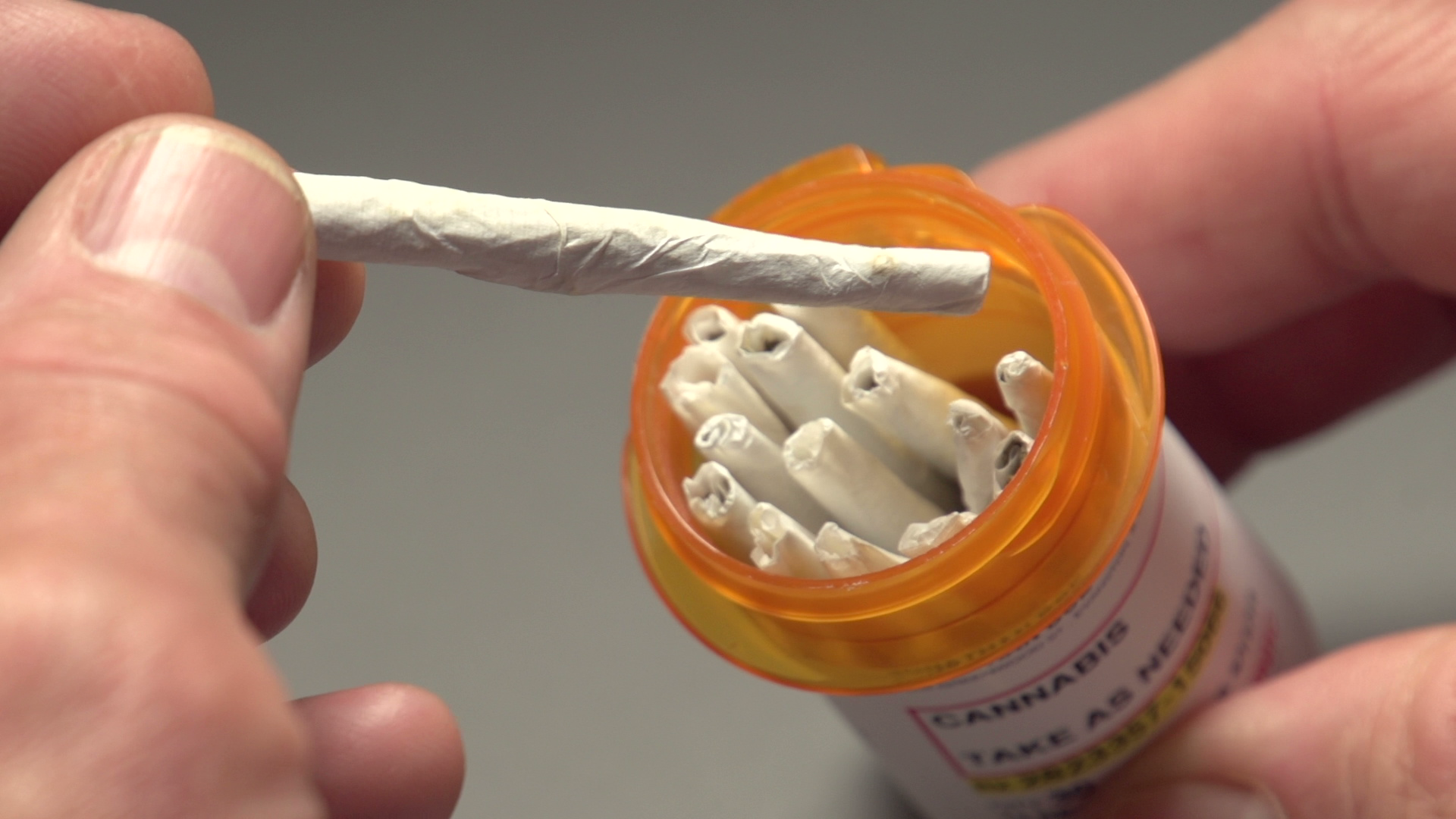
According to current trends, cannabis may soon be over prescribed and in many situations, unnecessarily so, yet the downside of that approach is less hazardous than the many toxic chemicals so casually dispensed.

Before you swallow beware! Instant gratification in the form of a pill is seldom a permanent solution and in some cases may be a deadly one.

Fred Kahn, MD, FRCS(C)

Antipsychotics, not anti-insomnia

National Post (Latest Edition)16 Jun 2017
Sharon Kirkey

Recently, after morning rounds seeing patients admitted to his hospital through emergency, Dr. David Juurlink tweeted: “Can the next doctor wanting to prescribe Seroquel for sleep, just not?”

Of the roughly 20 patients he had seen that morning, four had been prescribed Seroquel, an antipsychotic, for insomnia.

Seroquel and its generics aren’t approved as sleeping pills. Quetiapine, the active ingredient, has been officially approved in Canada for schizophrenia, bipolar disorder and major depression only.

Yet drug-safety experts are growing increasingly alarmed by the drug’s use as a doctor-prescribed nightcap for insomnia, with a 10- fold increase in quetiapine prescriptions for sleep problems in Canada between 2005 and 2012 alone.

Quetiapine is sedating. Like over- the- counter sleep aids, it makes people drowsy. But it also comes with a multitude of potential side effects, according to experts.

These side effects include an odd sensation of tension and restlessness (akathisia), Parkinson’s- like tremors and movement abnormalities, weight-gain, high blood sugar, new or worsening diabetes and, in rare cases, heart arrhythmia that can cause sudden cardiac death. A recent Health Canada review linked quetiapine and other so- called “atypical” antipsychotics to an increased risk of sleep apnea — breaks in breathing during sleep.

Juurlink, a clinical toxicologist at Sunnybrook Health Sciences Centre in Toronto, said quetiapine can also cause a particularly nasty complication known as neuroleptic malignant syndrome, a rare but potentially life- threatening reaction to antipsychotics or major tranquillizers. “Over the last decade, I have seen several patients who have had quetiapine as part of, or one of the contributing causes to NMS,” said Juurlink, whose frustrated tweet to doctors last week was a repeat of one he has sent before.

“I’ve certainly seen people who have been diagnosed with Parkinson’s disease that I’m confident were from quetiapine,” he added. “It’s getting to the point now where, when I admit a patient with Parkinson’s, I reflexively look at their other medications to see, ‘are they on quetiapine?’ ”

pills
According to drug market research firm IMS Brogan, of the 33 million prescriptions for tranquillizers dispensed by Canadian retail drugstores in 2016, one quarter — 8.3 million — were for quetiapine.

Doctors say the drug is being prescribed in low- dose formulations to people with no underlying psychiatric conditions, the majority for sleep. University of B.C. researchers found that 58 per cent of B.C. quetiapine prescriptions in 2010 were for the 25 mg tablet. The dose range for the approved disorders is 150 to 800 mg per day.

“It’s popping up as a patient’s typical medication for insomnia all the time,” says Kamloops emergency physician Dr. Ian Mitchell. “It’s not well supported by any science for use in sleep, it has significant side effects and yet it’s massively prescribed.”

“Seroquel is not benign,” Dr. David Gardner, a professor of psychiatry and pharmacology at Dalhousie University said in an email. “It may be more dangerous than our standard sleeping pills, but without research we cannot know or quantify its risks.”

It’s not clear how antipsychotics have become such a big thing for sleep. But observers point to aggressive marketing and industryfunded “opinion leaders” who’ve described quetiapine as a “mild, not harmful” drug that seems to help with sleep.

marijuana

Some users swear by it. “Seroquel helps me for sleep when nothing else will,” according to one online reviewer. “The only bad thing is 30 ( minutes) to one hour after taking it, I’m starving!!” Others describe feeling spacey and foggy the next morning.

Juurlink said quetiapine might shorten sleep latency — the time it takes to fully fall asleep — by a few minutes. It can also make people less aware of their “nocturnal awakenings” than they might otherwise have been. It’s a potent antihistamine, like diphenhydramine, the active ingredient in Benadryl and other “nighttime” cold remedies.

“But what’s really driving this is a societal expectation that we should all get eight hours of sleep a night, a pill is a way to go about it, and the willingness of some providers to accede to requests for sleeping pills,” Juurlink said.

While quetiapine has proven safe and effective for approved conditions, and most of the side effects have been reported during highdose treatment, side effects such as tardive dyskinesia — abnormal movements of the face and jaw — have been reported with low-dose regimens as well, according to the UBC Therapeutics Initiative.

Abuse of quetiapine is also a growing problem, with people inhaling or injecting crushed or dissolved tablets.

Mitchell says it’s hard to explain the “inherent hypocrisy” of the massive prescribing of an antipsychotic for insomnia, while medical leaders are warning doctors to be wary of prescribing marijuana for sleep.

“I’m not asking people to smoke a joint in a nursing home for sleep, that’s not what this is about,” he said. “But there may be some alternatives to some of the damaging medications that are out there, or ways to replace them with cannabis.”